
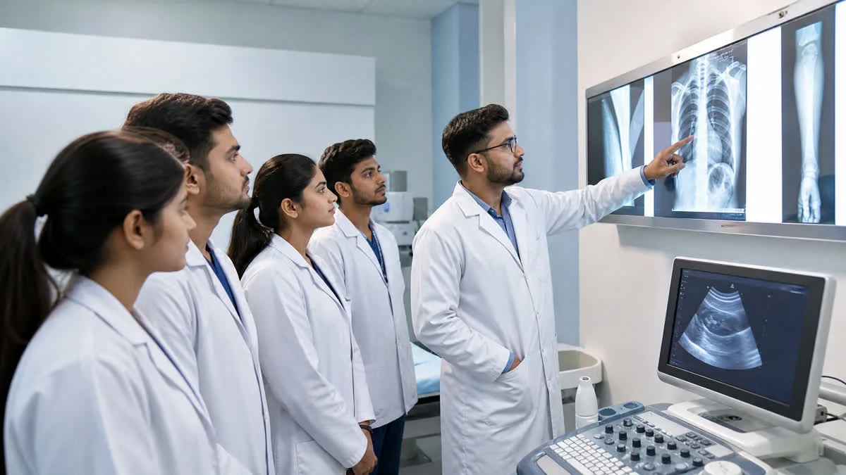
Medical Radiodiagnosis and Imaging Technology is the visionary eye of modern healthcare. Over 80% of all emergency trauma evaluations, neurological assessments, orthopaedic surgeries, and oncological stagings depend directly on high-precision radiological imaging. Radiologic Technologists operate cutting-edge diagnostic equipment—translating medical physics and human anatomy into diagnostic cross-sectional images that save lives every single day.
Philadelphia Mission Hospital Diagnostic Imaging Center • OSPMC Ambala. Below is the visual clinical protocol flowchart:
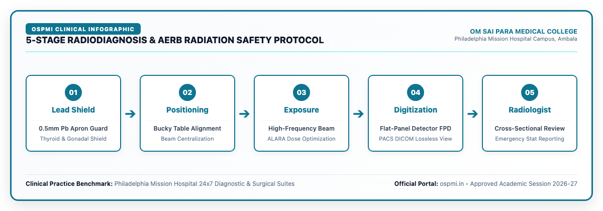
| Stage | Radiological Action | Equipment & Safety Tools | AERB Quality Benchmark |
|---|---|---|---|
| 01. Protection | Individual shielding of radiation-sensitive non-target anatomy | Lead rubber aprons (0.5mm Pb Eq), thyroid collars | > 95% attenuation of scattered radiation |
| 02. Positioning | Exact anatomical alignment relative to central X-ray beam | Motorized bucky grid table & vertical chest stand | Zero repeat-rate due to geometric cut-off |
| 03. Exposure | Precision calibration of kVp, mA, and automated exposure time | 500mA High-Frequency Generator with AEC chambers | ALARA compliance (< 20 mSv/year TLD monitor) |
| 04. Digitization | Sub-millimeter pixel matrix conversion & contrast tuning | Flat-Panel Digital Radiography (DR) & PACS Console | DICOM 3.0 lossless image transmission |
| 05. Diagnostic | Radiologist clinical interpretation & electronic dispatch | Medical-grade 5MP diagnostic monochrome monitors | Immediate emergency communication for trauma |
At Om Sai Para Medical College (OSPMC), Ambala, operating within the Philadelphia Mission Hospital campus since 2004, students receive rigorous clinical training on live diagnostic equipment, bridging classroom radiological physics with daily emergency hospital workflows.
The Evolution of Medical Imaging: From Roentgen to AI-Powered Multi-Slice CT
Ever since Wilhelm Conrad Roentgen discovered X-rays in 1895, diagnostic radiology has transformed medical science. Today's radiologic technologist does not merely take static photographic films. Modern medical imaging encompasses high-speed digital detectors, volumetric helical computed tomography, multi-parametric magnetic resonance imaging, and non-invasive vascular Doppler ultrasound.
Modern hospitals rely on technologists who understand digital matrix resolutions, signal-to-noise ratios (SNR), Hounsfield unit calibrations, and Picture Archiving and Communication Systems (PACS) that transmit diagnostic scans across global telemedicine networks in real time.
Core Diagnostic Modalities Mastered in RMIT
1. Digital Radiography (DR / CR Systems)
Digital radiography has completely replaced conventional wet chemical film processing. Technologists master:
- Flat Panel Detector (FPD) Technology: Direct and indirect digital conversion using cesium iodide (CsI) scintillators and amorphous silicon thin-film transistors (TFT).
- Specialized Contrast Examinations: Administering barium sulfate suspensions for Barium Swallow, Barium Meal follow-through, and Barium Enema; performing Intravenous Urography (IVU) and Hysterosalpingography (HSG) using water-soluble non-ionic iodinated contrast media.
- Skeletal & Trauma Positioning: Specialized projections including swimmer's view for cervicothoracic junction, scaphoid views, skyline patellar projections, and pediatric immobilization.
2. Computed Tomography (Multi-Detector CT-Scan)
CT scanning produces detailed cross-sectional tomographic slices of internal organs, eliminating tissue superimposition. Clinical training includes:
- Multi-Slice Spiral Acquisition: 16-slice, 64-slice, and 128-slice sub-millimeter volume scanning for polytrauma and stroke triage.
- CT Angiography (CTA): Power injector timing, bolus tracking techniques, and 3D volumetric vascular reconstruction for pulmonary embolism and aortic aneurysms.
- Artifact Management: Identifying and correcting motion artifacts, beam hardening, metal streak artifacts, and partial volume averaging.
3. Magnetic Resonance Imaging (MRI)
MRI provides unparalleled soft tissue contrast resolution without ionizing radiation by manipulating hydrogen proton spins in powerful superconducting magnetic fields (1.5 Tesla and 3.0 Tesla):
- Pulse Sequence Selection: T1-weighted imaging for anatomical definition, T2-weighted and FLAIR imaging for edema and demyelination, and Diffusion-Weighted Imaging (DWI) for hyperacute ischemic stroke detection within minutes of onset.
- Musculoskeletal & Spine Protocols: Evaluating cruciate ligament tears, meniscal pathology, intervertebral disc herniations, and spinal cord compressions.
- Strict MRI Safety & Zone Architecture: Managing Zone I through Zone IV safety perimeters, screening for pacemakers, cochlear implants, and ferromagnetic foreign bodies, and managing RF specific absorption rate (SAR) heating.
4. Radiation Safety & AERB Protection Standards
Patient safety and operator protection are the absolute cornerstones of radiological practice. Students at OSPMC are trained in strict compliance with the Atomic Energy Regulatory Board (AERB) guidelines and the international ALARA (As Low As Reasonably Achievable) principle:
- Cardinal Rules of Radiation Protection: Time minimization, distance maximization (inverse square law), and optimal structural shielding.
- Personal Protective Equipment (PPE): 0.5 mm lead-equivalent aprons, thyroid collars, lead acrylic mobile barriers, and gonad shields.
- Dosimetry Monitoring: Thermo-Luminescent Dosimeter (TLD) badge handling, quarterly dose evaluations, and occupational exposure thresholds.
Academic Pathways: Choosing Between Diploma, B.Voc & B.Sc
Students aspiring to enter radiodiagnosis can choose between three distinct recognized qualifications based on their career goals:
- Diploma in Radiology & Imaging Technology (DRIT - 2 Years): Focused on rapid job readiness, general radiography, darkroom techniques, and basic CT assistance. Ideal for students seeking swift employment in private clinics and district hospitals.
- B.Voc in Radiology & Medical Imaging Technology (3 Years): UGC-aligned skill degree offering 60% on-the-job hospital training with multiple exit points (Diploma after Year 1, Advanced Diploma after Year 2, Full Degree after Year 3). Perfect for students who want high clinical practical immersion and degree recognition.
- B.Sc. in Radiology & Medical Imaging Technology (4 Years): Comprehensive 4-year degree (3 Years Academic + 1 Year Hospital Internship) covering advanced radiological physics, research methodology, advanced MRI/CT post-processing, and eligibility for Master's degrees (M.Sc RMIT) and central government hospital recruitment.
Career Scope, Job Roles & Salary Trends in India & Abroad
Qualified radiology technologists enjoy exceptional job security across corporate hospitals, diagnostic imaging centers, and international healthcare systems:
- CT / MRI Technologist: Operating advanced multi-slice scanners in corporate chains (Max, Fortis, Medanta, Apollo). Average salary: ₹3.5 Lakh to ₹6.5 Lakh / year.
- Senior Radiographer (Government Sector): Permanent appointments in Civil Hospitals, AIIMS, ESI, and Railway Health Services via state public service commission examinations. Grade pay and allowances: ₹4.5 Lakh to ₹7.5 Lakh / year.
- Radiology Application Specialist: Working with multinational medical technology leaders like GE Healthcare, Siemens Healthineers, Philips, and Canon Medical, training hospital radiologists and technologists on newly installed scanners. Average salary: ₹6 Lakh to ₹10 Lakh / year.
- International Healthcare Opportunities (Gulf & UK): Massive global shortage of certified radiographers across the Middle East (UAE, Saudi Arabia, Qatar, Oman) and the UK National Health Service (NHS). Technologists clearing the DHA/HAAD or UK HCPC licensing examinations command tax-free salaries ranging from ₹12 Lakh to ₹22 Lakh / year.
Frequently Asked Questions (FAQs)
Q1: Is a career in Radiology dangerous due to radiation exposure?
No. When standard radiation safety protocols (AERB guidelines, lead shielding, collimation, and distance) are maintained, occupational exposure is virtually negligible and well within international safety limits.
Q2: Is NEET required for admission into Radiology courses?
No. Admissions to Diploma, B.Voc, and B.Sc Radiology programs at OSPMC Ambala are conducted through direct merit-based counselling based on your 10+2 marks.
Q3: Can a Radiologic Technologist start their own diagnostic center?
Yes. Qualified radiographers can establish registered digital X-ray and diagnostic sample collection facilities in compliance with local AERB radiation safety certifications and Chief Medical Officer (CMO) registrations.
Q4: Why choose OSPMC Ambala for Radiology training?
OSPMC operates directly inside the 22-year-old Philadelphia Mission Hospital campus in Ambala City, providing students with daily live patient examinations, digital bucky suites, and real emergency trauma scanning experience under senior radiologists.
Official 2026-27 Fee Structure and Installments
At Om Sai Para Medical College (OSPMC), Ambala, the annual tuition fee is payable in 3 installments every year:
• Diploma Programs (DMLT, OTT, Optometry, Dialysis, Cath Lab): ₹50,000 / year (₹15,000 + ₹17,500 + ₹17,500)
• B.Voc Degree Programs: ₹55,000 / year (₹15,000 + ₹20,000 + ₹20,000)
• B.Sc Degree Programs and BMLS: ₹60,000 / year (₹20,000 + ₹20,000 + ₹20,000)
• Admission form: ₹2,000 (one time). Examination fee: ₹1,000 per semester (Diploma, B.Voc), ₹2,000 per semester (B.Sc, BMLS)
0 comments
Be the first to comment on this article.